

Three-dimensional dental imaging gives dentists a complete spatial view of teeth, roots, nerves and bone that traditional flat X-rays simply cannot provide, allowing treatment decisions to be based on far more precise information. Instead of estimating depth and angle from a two-dimensional image, a dentist can rotate and examine a full 3D model of the exact area being treated, which changes how confidently and accurately many procedures can be planned.
Key takeaways
- 3D imaging captures depth and spatial relationships that flat X-rays cannot show.
- It allows more precise planning for implants, root canals and extractions.
- Nerve and sinus positions can be identified clearly before treatment begins.
- Fewer surprises during treatment often mean shorter, more predictable appointments.
- Radiation exposure is carefully managed and imaging is only used when it adds real value.
What Makes 3D Imaging Different From Traditional X-Rays?
Traditional dental X-rays compress a three-dimensional structure into a flat, two-dimensional image, which means depth information is essentially estimated rather than directly visible. Two structures that appear to overlap on a flat X-ray might actually be positioned at very different depths, and this ambiguity can make precise planning more difficult for complex cases.
3D imaging, often produced through cone beam computed tomography, captures hundreds of individual image slices that are then reconstructed into a detailed three-dimensional model. This model can be viewed from any angle, rotated, and examined slice by slice, giving a dentist the ability to see exactly how close a root sits to a nerve canal or how thick the bone is at a specific point, rather than relying on trained estimation from a flat image.
How Does This Improve Planning for Dental Implants?
Placing a dental implant successfully depends on accurately understanding the width, height and density of the jawbone at the exact planned location. 3D imaging allows a dentist to measure these dimensions precisely before any surgery begins, identifying whether there is sufficient bone or whether additional preparation might be needed first.
This level of detail also allows the exact angle and depth of implant placement to be planned in advance, sometimes using the 3D model to create a surgical guide that helps position the implant with high accuracy during the procedure itself. This reduces guesswork during surgery and helps ensure the implant is positioned in a way that supports both function and long-term stability.
Why Is Identifying Nerve and Sinus Position So Important?
Certain structures in the jaw, particularly the nerve canal in the lower jaw and the sinus cavities above the upper back teeth, need to be avoided during many dental procedures. On a flat X-ray, judging exactly how close these structures are to a treatment site can be difficult, especially in cases where anatomy varies from what is typical.
3D imaging shows the precise position of these structures relative to the treatment area, allowing a dentist to plan around them with far greater confidence. This is particularly valuable for lower back tooth extractions or implants, where nerve proximity is a common concern, and for upper back teeth procedures where sinus position needs to be considered carefully.
How Does 3D Imaging Support More Complex Root Canal Cases?
Root canal treatment depends on locating and cleaning every canal within a tooth’s root system, but root anatomy can be unpredictable, with extra canals, unusual curves or narrow branching that are not always visible on a standard X-ray. Missing a canal during treatment can affect the long-term success of the procedure.
With 3D imaging, a dentist can examine the internal root structure of a specific tooth in detail before treatment begins, identifying the number, shape and curvature of canals present. This allows a more accurate and complete approach to be planned from the outset, particularly useful for teeth known to have complex or atypical root anatomy.
What Difference Does This Make for Complicated Extractions?
Some extractions, particularly of impacted wisdom teeth, involve teeth positioned at difficult angles or close to sensitive structures. Understanding the exact orientation of the tooth, its roots, and its relationship to nearby nerves before beginning the procedure allows a more informed and controlled approach.
3D imaging lets a dentist see whether roots are curved in a way that might complicate removal, or whether a tooth’s position places it unusually close to a nerve canal. This information can influence decisions about technique, and in some cases whether referral to a specialist is the more appropriate path, all determined before the appointment rather than discovered partway through.
Does 3D Imaging Reduce Treatment Time and Surprises?
When a dentist has a complete, accurate picture of the treatment area beforehand, there is less need to pause during a procedure to reassess an unexpected finding. This generally translates into more predictable appointment lengths and fewer situations where a procedure needs to be adjusted or rescheduled once it is already underway.
Patients often experience this as smoother, more efficient visits, since much of the uncertainty that might otherwise arise during treatment has already been addressed during planning. While no imaging technology eliminates all variability in individual healing or anatomy, thorough pre-treatment planning meaningfully reduces the likelihood of surprises.
How Is Radiation Exposure Managed With 3D Imaging?
Cone beam 3D imaging generally involves more radiation than a single standard dental X-ray, though modern equipment is designed to keep doses as low as reasonably possible while still producing useful detail. Dentists weigh the diagnostic value of a 3D scan against this exposure, using it selectively rather than as a routine replacement for standard X-rays.
This selective approach means 3D imaging is typically reserved for cases where the additional detail meaningfully changes treatment planning, such as implants, complex extractions or unusual root anatomy, rather than being used for routine checkups where a standard X-ray already provides sufficient information.
What Should Patients Expect During a 3D Scan?
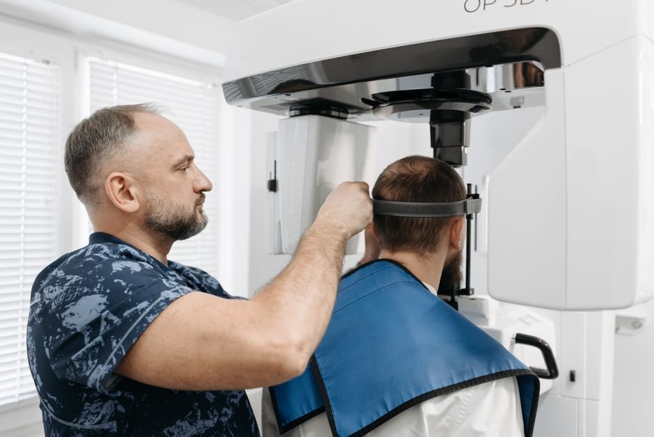
A 3D dental scan is generally quick and non-invasive, often taking less than a minute of actual scanning time while a machine rotates around the head to capture the necessary images. Patients typically remain still in a seated or standing position, and no injections or preparation are usually required beforehand.
Afterwards, the images are processed into a three-dimensional model that the dentist can review, often within the same appointment. This allows treatment discussions to happen with the detailed imagery already available, giving patients a clearer picture of their own anatomy and the reasoning behind a proposed treatment plan.
How Does This Technology Support Better Patient Communication?
Beyond planning benefits, 3D imaging gives dentists a visual tool that can help explain a proposed treatment in a way flat X-rays often cannot. Being able to show a patient a rotatable model of their own jaw, pointing to the exact area of concern, tends to make explanations clearer and more concrete.
This visual clarity can make it easier for patients to understand why a particular treatment approach is being recommended, what risks are being carefully planned around, and what the procedure will involve. Clearer communication at the planning stage often contributes to patients feeling more informed and comfortable going into treatment.
Frequently Asked Questions
Is 3D imaging used for every dental procedure?
No, 3D imaging is generally reserved for cases where the additional detail meaningfully changes treatment planning, such as implants, complex extractions or root canals with unusual anatomy. Routine checkups and simple procedures typically continue to use standard dental X-rays, which provide sufficient information for those situations.
Does a 3D scan take longer than a regular dental X-ray?
The actual scanning process is usually quick, often completed in under a minute, though it may take slightly longer than a single standard X-ray image. The greater time investment is typically in processing and reviewing the resulting three-dimensional model rather than the scan itself.
Is 3D imaging safe for regular use?
3D imaging involves a higher radiation dose than a single standard X-ray, which is why it is used selectively rather than routinely. Dentists follow guidelines to ensure imaging is only used when the diagnostic benefit clearly outweighs the exposure, keeping overall use appropriate and justified.
Can 3D imaging detect problems that regular X-rays miss?
Yes, because it captures depth and spatial detail that flat X-rays cannot show, 3D imaging can reveal issues such as hidden extra root canals, precise nerve proximity, or subtle bone density variations that might not be clearly visible on a standard two-dimensional image.
Does insurance typically cover 3D dental imaging?
Coverage varies depending on individual plans and the reason the imaging is being performed. It is generally worth checking directly with a dental provider or insurer about coverage for a specific procedure, since policies differ on when advanced imaging is considered necessary versus optional.