


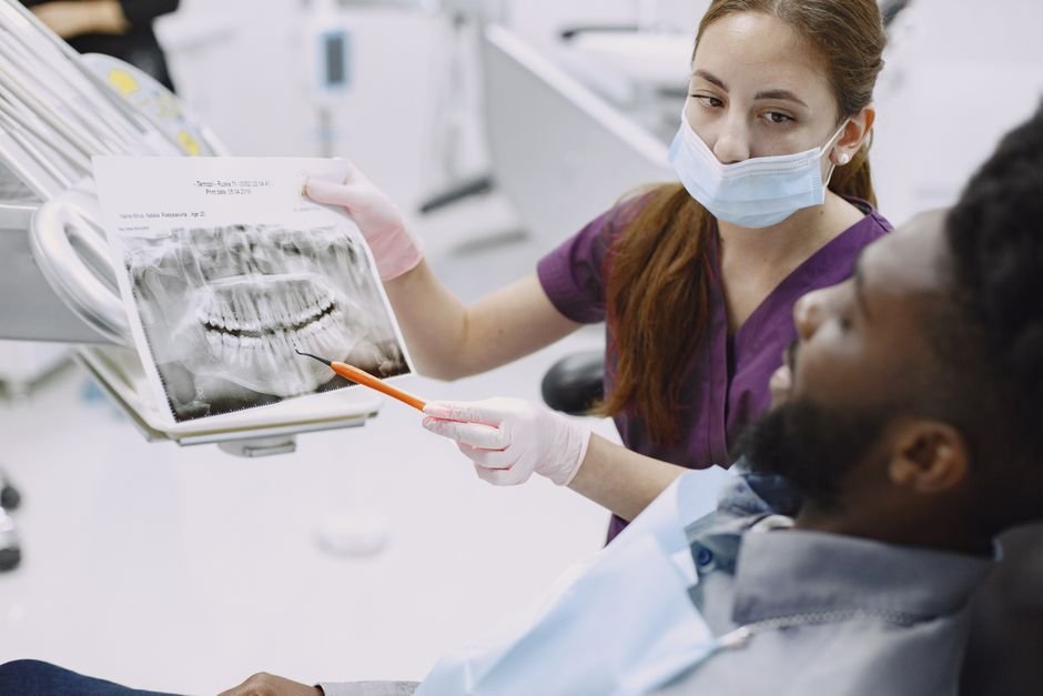
Dental X-rays are imaging tools that let a dentist see the parts of your teeth and jaw that are hidden beneath the gums and inside the tooth structure itself. They reveal decay between teeth, bone loss, infections at the root tip, impacted teeth, and other problems long before they would ever become visible or painful. In short, they turn a visual check-up into a far more complete picture of your oral health, allowing small issues to be caught and treated while they are still simple and inexpensive to manage.
Key takeaways
- Dental X-rays show decay, bone loss, and infections that cannot be seen during a visual examination alone.
- Modern digital X-ray equipment uses very low levels of radiation, comparable to everyday background exposure.
- How often you need X-rays depends on your individual risk of decay and gum disease, not a fixed schedule for everyone.
- Protective aprons and thyroid collars are used routinely to minimise any radiation reaching the rest of the body.
- Skipping X-rays for long periods can allow hidden problems to progress silently until they become more serious and costly to treat.
What Are Dental X-Rays and Why Are They Used?
A dental X-ray is a two-dimensional image created by passing a small, controlled dose of radiation through the teeth and jaw onto a sensor or film. Because dense structures such as enamel and bone absorb more radiation than softer tissue, they appear lighter on the resulting image, while areas of decay, infection, or bone loss appear darker. This contrast allows a dentist to identify subtle changes that would otherwise remain completely invisible during a standard visual check-up.
Without X-rays, a dentist can only assess what is visible on the surface of the teeth and gums. Many common problems, however, begin in places the eye simply cannot reach, such as between tightly spaced teeth or underneath old fillings. X-rays close this gap in information, giving the dental team a much fuller understanding of what is actually happening inside the mouth, which in turn supports more accurate diagnosis and more conservative, timely treatment.
How Do Dental X-Rays Work?
The process relies on the fact that different tissues in the body absorb X-ray radiation to different degrees. Teeth and bone are dense and absorb a large proportion of the radiation, so they appear as lighter areas on the image. Softer tissues, decayed portions of a tooth, and the spaces around the roots absorb less radiation and therefore show up as darker regions. A sensor placed inside or against the mouth captures this pattern and converts it into an image the dentist can study closely.
Most practices now use digital sensors rather than traditional film, which means the image appears on a screen within seconds rather than requiring chemical development. Digital systems also allow the image to be enlarged, adjusted for contrast, and compared side by side with earlier scans, making subtle changes over time much easier to track. This shift to digital technology has also considerably reduced the radiation dose needed to produce a clear, diagnostic image.
What Types of Dental X-Rays Are There?
Several different types of dental X-ray exist, each suited to a particular purpose. Bitewing X-rays capture the upper and lower back teeth in a single image and are especially useful for spotting decay between teeth and checking the fit of existing fillings. Periapical X-rays show a complete tooth from crown to root tip, which helps identify infections, abscesses, or unusual root shapes. Panoramic X-rays capture the entire mouth, including the jaw, sinuses, and any unerupted teeth, in one wide image.
More advanced imaging, such as three-dimensional cone beam scans, may be used for complex cases like planning dental implants or assessing impacted wisdom teeth in fine detail. The choice of which type to use depends entirely on what the dentist needs to evaluate. A routine check-up might only call for bitewings, while planning a more involved procedure could require a panoramic or three-dimensional image to give a complete surgical picture.
How Often Should You Have Dental X-Rays Taken?
There is no single schedule that applies to everyone, because the right frequency depends on individual risk factors. Someone with a history of frequent cavities, gum disease, or previous dental work may need X-rays more often than someone with consistently healthy teeth and low decay risk. Age, oral hygiene habits, and whether orthodontic treatment is underway can all influence how regularly imaging is recommended, so the interval is always tailored rather than fixed.
For many adults with good oral health and no ongoing concerns, X-rays every one to two years is a common approach, while children and teenagers, whose teeth and jaws are still developing, may need them more frequently to monitor growth and eruption patterns. A dentist will review your history at each visit and decide whether new images are genuinely needed, rather than taking them automatically at every appointment regardless of circumstance.
Are Dental X-Rays Safe?
Dental X-rays involve an extremely small dose of radiation, and modern digital equipment has reduced this further compared with older film-based systems. To put it in perspective, the radiation from a typical set of bitewing X-rays is roughly comparable to the background radiation a person is naturally exposed to over the course of a single day simply from the environment around them. This makes the individual risk from any one dental X-ray very low for the vast majority of patients.
Dental teams also follow strict protocols to keep exposure as low as reasonably possible. Lead-lined aprons and thyroid collars are commonly used to shield the rest of the body, and equipment is positioned carefully so that only the specific area being examined is captured. Anyone with particular concerns, including those who are pregnant, should always mention this beforehand so the dental team can discuss the safest approach or postpone non-urgent imaging if appropriate.
What Can Dental X-Rays Reveal That a Visual Exam Cannot?
A visual examination, even a very thorough one, only shows the outer surfaces of the teeth and gums. X-rays reveal what lies beneath, including decay hiding between teeth where a probe or mirror cannot reach, infections forming at the tip of a tooth root, and bone loss around the teeth that signals advancing gum disease. They can also detect problems with teeth that have not yet broken through the gum line, such as impacted wisdom teeth.
Beyond decay and infection, X-rays help identify cysts, unusual growths, and changes in the jawbone that would otherwise go unnoticed until they caused pain or visible swelling. They also allow a dentist to check the condition of previous dental work, such as whether a filling or crown still fits correctly or whether a root canal has healed as expected. This hidden information is often the difference between catching a problem early and only discovering it once it has become far more extensive.
What Happens During a Dental X-Ray Appointment?
Taking a dental X-ray is generally quick and straightforward. For most images, you will be asked to bite down gently on a small sensor or piece of film positioned inside the mouth, or to hold still while a larger machine rotates around your head for a panoramic image. A protective apron is placed over your chest, and sometimes a collar is added to shield the thyroid area, before the image is captured in a matter of seconds.
There is no discomfort involved, though some people find the sensor slightly awkward to bite on, particularly if they have a strong gag reflex or a smaller mouth. The dentist or dental nurse will explain what is happening at each step and can adjust the positioning if something feels uncomfortable. Once captured, the image typically appears on a screen almost immediately, allowing the dentist to review it with you as part of the same appointment.
Common Myths About Dental X-Rays
One persistent myth is that dental X-rays carry a significant radiation risk, when in reality the dose involved is very small and tightly regulated. Another common misconception is that X-rays are only needed when a problem is already suspected, but their real value lies in detecting issues before any symptoms appear, when treatment is simplest and least invasive. Waiting for pain before agreeing to an X-ray often means a problem has already progressed further than it needed to.
Some people also believe that if their teeth look and feel fine, X-rays are unnecessary altogether. However, many of the conditions X-rays detect, including early decay between teeth and the initial stages of bone loss from gum disease, produce no noticeable symptoms until they are well advanced. Regular imaging, at intervals appropriate to individual risk, remains one of the most reliable ways to catch these silent changes while they are still easy to address.
How Should You Prepare for a Dental X-Ray?
Preparation for a routine dental X-ray is minimal. There is no need to fast, avoid food or drink, or make any special changes to your daily routine beforehand. The main practical step is simply removing any metal objects that could interfere with the image, such as earrings, necklaces, or a removable retainer, since metal can create shadows or artefacts on the resulting picture and reduce its clarity.
It is worth mentioning any relevant medical information to the dental team before imaging begins, particularly pregnancy or any recent radiation exposure from other medical procedures, so this can be factored into the decision. Letting the team know in advance if you have a sensitive gag reflex or find certain positions uncomfortable also helps, as adjustments can often be made to the equipment or technique to make the process considerably more comfortable.
Frequently Asked Questions
Do dental X-rays hurt?
No, taking a dental X-ray is not painful. You may feel mild pressure from biting on a sensor or holding a positioning device in place, but the process itself involves no discomfort and is completed within seconds.
Can dental X-rays detect gum disease?
Yes, X-rays are particularly useful for assessing gum disease because they show the level of bone supporting the teeth. A dentist can compare bone height over time to track whether gum disease is stable, improving, or progressing beneath the surface.
How long does it take to get dental X-ray results?
With digital equipment, images typically appear on screen within seconds of being taken. The dentist can usually review and discuss the results with you during the same appointment, rather than requiring a separate visit.
Are children’s dental X-rays different from adult ones?
The underlying technology is the same, though the frequency and type of imaging may differ. Children’s X-rays often focus on monitoring developing teeth, jaw growth, and the position of teeth that have not yet erupted.
What happens if you refuse dental X-rays?
A dentist can still perform a visual examination, but without X-rays, decay between teeth, hidden infections, and early bone loss may go undetected. This can mean problems are only found once they have grown larger, more painful, and more complex to treat.